Arteriovenous Malformation AVM

Arteriovenous Malformation

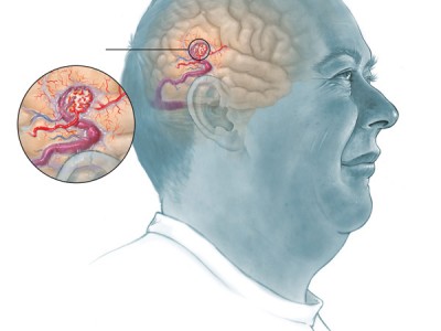
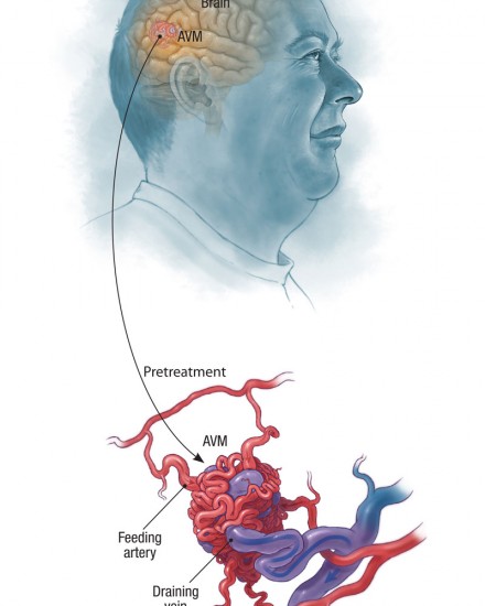
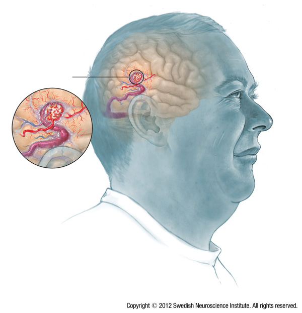
Arteriovenous malformation (AVM) is a tangled group of blood vessels with abnormal connections between arteries and veins. AVMs are rare — affecting less than 0.1 percent of Americans.

An AVM can occur in the brain or spinal cord, and are particularly dangerous in the brain. They can lead to headaches, seizures, or bleeding into the brain as well as stroke-like symptoms and, in rare cases, the risk of a fatal stroke.

AVMs are found in people of both sexes and all racial and ethnic backgrounds, either on scans of the brain or spinal cord, or after they have caused symptoms. AVMs form in the fetus during pregnancy, but their cause is unknown. They are not associated with any activity or medication during pregnancy or any complications of birth. Dr Newell has been treating AVM’s for over 25 years using surgical techniques as well as Gamma knife radiosurgery. He has extensive surgical skills used to remove ruptured and unrupured AVM’s.

Symptoms

Most people with AVMs have no symptoms. Often, they are found when patients are screened for another condition, and an AVM becomes visible on a CT scan or an MRI.

Symptoms do occur in about 12 percent of people with AVMs. These are typically seen between the ages of 10 and 30 and can include:

  • Sudden and severe headaches or seizures
  • Sudden muscle weakness or paralysis
  • Numbness and tingling
  • Problems with vision, language use, coordination, or memory

What are the risks?

The greatest potential risk to someone with an AVM is bleeding in the brain. Because the smaller blood vessels in an AVM are abnormal, they may leak or rupture.

When high pressure causes an AVM to rupture, a large, rapid accumulation of blood – called a hemorrhage – can occur. About 2 to 4 percent of all AVMs each year result in hemorrhages.

Hemorrhaging from a ruptured AVM can cause stroke-like symptoms or a fatal stroke. Young people between the ages of 15 and 20 who are diagnosed with AVMs are at the highest risk of hemorrhaging.

Patients with an AVM are carefully monitored for any signs that may indicate an increased risk of hemorrhage. If a hemorrhage occurs and is large, the AVM needs to be surgically removed immediately.

During treatment for AVMs, a risk of hemorrhage remains until the AVM is completely gone.

Factors Affecting AVMs

Once formed, various external factors may affect the development, size and shape of AVMs.

Arterial Shunting

Normally, before a baby is born and starts using the lungs to breathe, blood from the heart headed for the lungs in the pulmonary artery is shunted (diverted) away from the lungs and returned to the heart. Other arteries become smaller and smaller as they reach the tissues they supply. Then they break into capillaries and finally they drain into veins.

When there is direct arterial shunting from the artery to the vein, the result can be a tangle of vessels that make up an AVM. This shunting causes low blood pressure in the arterial vessels feeding the AVM and neighboring areas of the brain that they normally supply with blood.

Intracranial Hemorrhage

An intracranial hemorrhage is bleeding within the cranium (skull) which can be caused by an AVM. This can also be caused by a stroke or an aneurysm in the brain. Abnormal changes in blood pressure make AVMs vulnerable to causing an intracranial hemorrhage.

Syndromes that cause AVMs

There are a small number of syndromes that people have from birth that are cause AVMs. These syndromes are very rare.

Sturge–Weber syndrome results in the deformity of blood vessels on the surface of the brain. In rare cases it may cause a severe form of epilepsy, mental retardation or progressive shrinkage of the brain.

Osler-Weber-Rendu syndrome is a genetic disorder that leads to the development of abnormal blood vessels in the skin, mucous membranes, and often in the lungs, liver or brain. Patients with Osler-Weber-Rendu syndrome usually suffer from constant nosebleeds or strokes.

Treatment

The goal of treatment is to prevent complications by controlling the risk of bleeding and seizures and, if possible, to remove the AVM. It is important to complete a thorough diagnostic workup to ensure an accurate evaluation. For example, an MRI may allow the surgeon to determine if an AVM is near critical brain areas.

A treatment plan is then developed for each patient, based on a number of factors, including:

  • The size of the AVM
  • Its location and the blood vessels involved
  • A person’s age and medical history

The neurosurgeons and endovascular surgeons  use state-of-the-art technology to perform a number of different treatments including microsurgery, embolization, and radiosurgery to treat AVMs.

Following treatment, imaging tests are repeated to see if the AVM was completely removed, or if additional treatment is needed. Patients are monitored closely for completion of the treatment plan with ongoing visits and additional imaging testing if needed.

Microsurgery

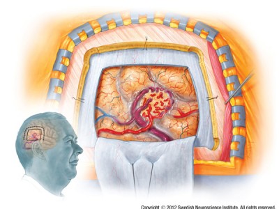
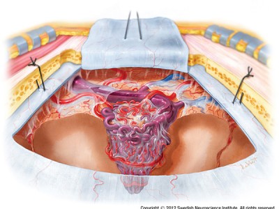
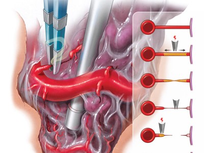
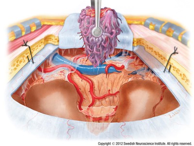
Microsurgery involves special techniques to close off the blood vessels that are feeding the AVM and remove the AVM, which in turns removes the risk of future bleeding. The image below is an example of techniques used by the neurosurgeon  to remove an AVM.

Embolization

Embolization, also known as selective vessel AVM embolization, is an interventional radiology procedure in which a special surgical “glue” is inserted into the AVM to block the blood flow. Under X-ray guidance, a very thin tube called a catheter is inserted into the femoral artery and threaded up into the brain. This surgical “glue” is then delivered to a specific area in the brain in order to block the blood flow to the AVM. 

This procedure may be done as one procedure, or as part of a staged procedure. Embolization is performed by an endovascular surgeon.

Radiosurgery

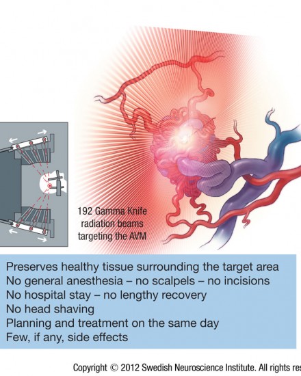
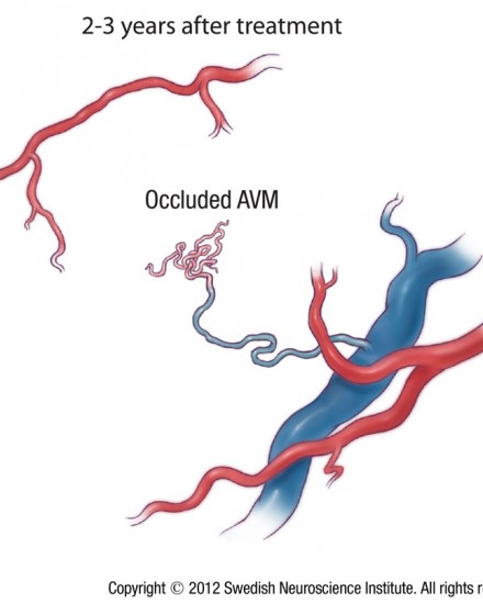
Radiosurgery involves delivering of a high dose of radiation directly to an AVM. This causes the blood flow to slowly close off over a period of time until it eventually stops. This treatment is often selected if AVMs are in deep locations in the brain, making surgery difficult.

Radiosurgery is also used when the AVM is too close to regions of the brain that are critical to everyday living including speech, motor and visual areas. The team at our Center utilizes the Perfexion™ Gamma Knife — which is the most proven and versatile radiosurgery equipment available for AVM treatment.

Video

David Newell, M.D. gives an overview of arteriovenous malformations or AVMs, which are a collection of blood vessels that can occur anywhere within the brain or spinal cord.

Arteriovenous Malformation AVM. For help with symptoms, risks, and treatment from our world renowned doctors contact us or visit us at Seattle Neurosciences’ website.

Dr. Newell was born in Boston, MA and attended Case Western Reserve University medical school. He then completed his residency in neurosurgery at the University of Washington, including one year in London at St. George’s medical school. Dr. Newell is the co-founder of the Swedish Neuroscience Institute and founder of the Seattle Neuroscience Institute.

If you need help with any of these conditions, please contact us to get help!

Or call 206.661.6100 for immediate assistance